Information for families

You’re expecting twins, now what?

The Twins Research Centre strives to empower and educate families about their twin pregnancy, labour and delivery. In this area, we’ll provide an overview of common terms you will hear, as well as some common questions and address concerns you may have with your twin pregnancy.

Pregnant woman doing research
General information

Learning a new language: Common medical terms

Twin pregnancies can be described or classified in two ways. The first is in relation to zygosity (the initial number of fertilized eggs that resulted in a twin pregnancy), and the second is in relation to placentation (the number of placentas and amniotic sacs).


  1. Zygosity

Zygosity refers to the genetic relationship between your twins. It stems from “zygote,” which refers to the single fertilized egg. There are two types of zygosity:

  • Monozygotic twins, also known as identical twins, form when a single fertilized egg splits, resulting in the development of two individual embryos. Because they originate from the same set of cells, these individuals have the same DNA and often have remarkably similar physical appearances.
  • Dizygotic twins, also known as fraternal twins, form when multiple eggs are fertilized and develop. They may look alike or may look completely different, the same as any siblings would.


Opposite-sex twins (male and female) are always dizygotic. However, in the case of same-sex twins, determining whether they are identical (monozygotic) or non-identical (dizygotic) may require additional information, including ultrasound findings regarding chorionicity (as described below), blood type, or DNA analysis.


  1. Placentation

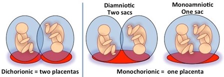
Placentation refers to the number of placentas (or chorionicity) and amniotic sacs (or amnionicity) that are present in the twin gestation.


Chorionicity

Chorionicity refers to whether each of your babies has their own placenta (known as dichorionic twins) or whether they share one placenta (monochorionic twins). It’s important to find out early on during your pregnancy whether your babies share a single placenta since this puts you and your babies at a higher risk of complications.


Amnionicity

Amnionicity refers to whether each of your babies is located within their own inner fluid-filled sac (known as diamniotic twins) or whether they share a single sac and thus have no membranes separating them (monoamniotic twins).


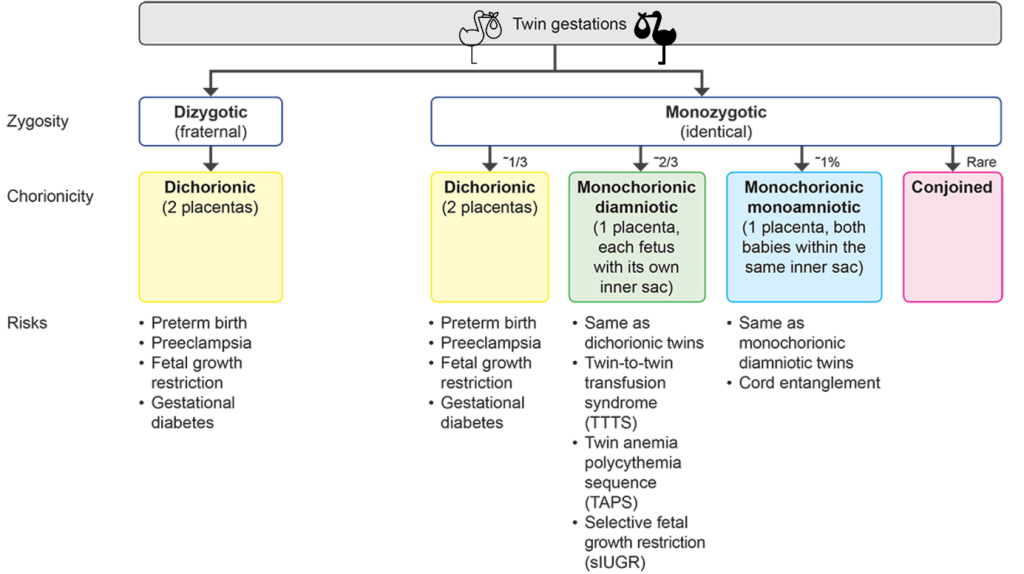
Relationship between zygosity, chorionicity, and amnionicity


Source: The Johns Hopkins Center for Fetal Therapy

Dizygotic (fraternal) twins would always be dichorionic and diamniotic, since each twin originates from a separate fertilized egg, each developing into a fetus with its own membranes and placenta.


In contrast, monozygotic (identical) twins may share the placenta or amniotic sac, depending on when the single zygote splits, since the two babies will have to share all the structures that developed before the split occurs. Thus, if the egg splits at a very early stage (usually within the first three days following fertilization, before the placenta begins to form), each baby will have its own placenta and amniotic sac, which results in dichorionic diamniotic twins.  If the egg splits at a later stage (between days 3-8, when the placenta has already begun to form but the amniotic sac has not), the resulting twins will share the same placenta, resulting in monochorionic diamniotic twins. If the split happens even later (between days 8-13, at which time the amniotic sac was already formed), the two babies will share not only the placenta but also a single sac, resulting in monochorionic monoamniotic twins. Finally, in the rare event that the egg splits at an even later stage (after day 14, when the baby’s body has already started to develop), the two twins will share not only the same placenta and amniotic sac but also the body parts that were already formed at the time of the split, resulting in conjoined twins.


Both care providers and couples with twins occasionally get confused and assume that dichorionic twins (who have 2 separate placentas) are always non-identical (i.e., dizygotic). However, as described above and seen in the figure below, this is not always the case, and about a third of identical (monozygotic) twins will be dichorionic.


Epidemiology of twins

Twin pregnancies account for 1-2% of overall pregnancies. The incidence of triplets (and higher-order multifetal pregnancies such as quadruplets) is much lower, at approximately 0.08%.


The incidence of twin pregnancies is calculated by the number of twin pregnancies out of overall pregnancies per a given time period. It can also be estimated by Hellin’s Law (named after the German statistician Wilhelm Hellin), which is a mathematical formula. It says that for every 89 singleton prengnacies, there will be 1 twin pregnancy (1:89 ratio), for every 89 twin pregnancies there will be 1 triplet pregnancy etc.


In Canada, about 6000 sets of twins are born each year. In Sunnybrook, we take care of about 350 mothers with twins every year.


You are at increased risk of having a twin pregnancy if you:

  1. Used assisted reproductive technology (i.e., in-vitro fertilization (IVF), ovulation induction, superovulation plus intrauterine insemination) – Ovulation-inducing agents increase the chance that multiple eggs are released and then fertilized, resulting in dizygotic twins. In addition, IVF has also been shown to increase the risk of monozygotic twins. It is believed that the fact that the embryos are initially grown in a culture dish (as opposed to within the uterine cavity) might increase the chance that the single fertilized egg will split.
  2. Are older (over 35 years): Why this is true is not well-known, but it is believed that older mothers are more likely to release multiple eggs due to different hormone levels secreted from their ovaries (e.g., inhibin) compared to younger mothers.
  3. Have a personal or family history of twin pregnancies: there is a well-established genetic predisposition to having dizygotic twins. This predisposition has been linked to specific genes that affect the levels of hormones or the response to hormones (e.g., the follicle-stimulating hormone [FSH]receptor) involved in the ovulation process. This genetic predisposition also explains the high rate of twin pregnancies in certain countries (e.g., Nigeria).
  4. Are tall or overweight: Tall mothers (over 164 cm) and obese mothers (BMI of 30 or greater) have a higher chance of a twin pregnancy.

Why am I considered to have a ``high risk`` pregnancy?

When you’re carrying twins, you’re at greater risk for a number of complications. Because of this, you will be followed by an obstetrician or a special ‘high risk’ obstetrician called a “maternal fetal medicine specialist” (MFM). You are at a higher risk for:

  • Premature birth – this means delivering your twins before the end of 36 weeks of pregnancy. This happens in roughly half of all twin pregnancies. Premature babies may have more health problems.
  • Low birth weight – More than half of twins are born weighing less than 5 1/2 pounds, which is considered “low birth weight” (LBW). Babies born at a low birth weight are at increased risk of health problems after birth, such as vision and hearing problems, mental disabilities, and cerebral palsy (with the risk greatly increasing if babies are born before 32 weeks or weighing less than 3 1/3 pounds).
  • Twin-twin transfusion syndrome (TTTS) – This condition develops when a connection between the babies’ blood vessels allows one baby to get too little blood and the other too much. This complication is unique to monochorionic twins (who have a single placenta), and develops in approximately 10-15% of these pregnancies. A doctor can treat TTTS with laser surgery to seal off the vessel connection or with amniocentesis to drain excess amniotic fluid.
  • Preeclampsia – This is a pregnancy complication characterized by high blood pressure and signs of damage to the liver and/or kidneys. You may have swelling, headaches, and vision changes, in addition to the high blood pressure. If not treated, preeclampsia can deprive your baby of oxygen and nutrients, and can also damage your organs.
  • Gestational diabetes – This condition means developing diabetes during your pregnancy and may result in your babies growing too big. Delivering a large baby increases your risk of labour and delivery complications, and your babies may have breathing problems and low-blood sugar when they’re born.

This all may sound pretty scary, but the good news is that you and your health care team can lower your risk and identify any problems earlier, rather than later. You can then work with your health care providers to manage and minimize the impact of any complications. You can read more about all of these conditions below.

Risks of twin pregnancies

Preterm birth

About 60% of twin babies will be born preterm (before 37 weeks of gestation). Being born early, especially before 32 weeks, can result in both short- and long-term complications. It’s not clear why twin babies are born early, but it’s believed to be, at least in part, due to the over-distension (or extra stretching) of the uterus.


Epidemiology

Below are some statistics adapted from Martin JA et al. (2021) showing the incidence of preterm birth and preeclampsia, as well as birthweight and gestational age at birth, by type of pregnancy.

Outcome  Singletons Twins Triplets
Preterm birth<37 weeks 8% 60% 99%
Preterm birth <34 weeks 2% 15-20% 63%
Preterm birth <28 weeks 4%
Average weeks of gestation at birth 39 35-37 32-33 (quadruplets 30w)
Average birthweight 3286g (7.2 lbs) 2345g (5.2 lbs) 1681g (3.7 lbs)
Birthweight <2500g (5.5 lbs) 6% 55% 95%
Birthweight <1500g (3.3 lbs) 1% 9% 34%
Preeclampsia 4% 8% 10%


Risks of preterm birth

Preterm babies are at higher risk of short- and long-term complications. At birth, they may have breathing and feeding difficulties, be at higher risk of infection or visual impairment, and have severe neurological and gastrointestinal complications. In the long term, they may experience growth delay or neurodevelopmental impairment.


Diagnosis

Preterm birth feels just like a regular birth, just early! Some things to look out for are contractions (which may feel like abdominal cramping, tightening, or period-like lower back pain), pelvic pressure, leakage of clear (amniotic) fluid, vaginal bleeding, or increased vaginal discharge. You should speak with your doctor or visit the hospital if you experience these symptoms.


Management of preterm birth

A mother’s cervix has to shorten and dilate during birth to allow for the baby’s head to pass from the uterus into the vaginal canal. Sometimes, the cervix shortens or dilates too early, a finding that increases the risk of preterm birth. At Sunnybrook’s Twins Clinic, we will measure your cervix via vaginal ultrasound every 2 weeks between 16 and 28 weeks gestation to detect if it is shortening or dilating. This allows us to detect such changes early on (before you experience any symptoms) and take actions that may decrease the risk of early delivery, such as the administration of vaginal progesterone (if the cervical length is below 25mm), or the use of a stitch to strengthen the cervix (known as cervical cerclage, if the cervical length is less than 15mm or the cervix is open).

Once preterm birth has already started, it cannot be stopped, but it can be slowed down to maximize the health of your babies. Drugs called tocolytics can be given to slow uterine contractions, allowing for more time to give your babies medications that help them thrive after birth. Examples are antenatal corticosteroids, which accelerate fetal lung development. If delivery is imminent, we may give you magnesium sulphate, which is a medication that helps protect the baby’s brain.

Fetal growth restriction

Fetal growth restriction (FGR) is defined as a failure for the baby to meet its growth potential due to a pathological process, most commonly a failure of the placenta to support fetal growth (known as placental insufficiency). In monochorionic twin pregnancies, a specific type of FGR occurs called selective fetal growth restriction (sFGR), which is discussed in more detail below (section 5).


Epidemiology

In singleton pregnancies, up to 10% of babies may be diagnosed with FGR. Twin babies grow slower than singletons starting at around 26-28 weeks of gestation. As a result, up to 30-50% of twin babies would be identified as small (and potentially growth-restricted) when their growth is evaluated using singleton growth charts. It is unclear, however, whether the slower growth of twins is due to a truly higher incidence of FGR due to placental insufficiency, or whether this is a benign physiologic adaptation of twin babies meant to reduce uterine over-stretching and, consequently, the risk of preterm birth.

More in the How is FGR in twin pregnancies different than in singleton ones?’ section below!


Risks of FGR

Short term, FGR babies are at risk of mortality and morbidity, including respiratory, neurological, infectious, and gastrointestinal complications. Long-term complications of FGR include neurodevelopmental delay and increased risk of developing heart disease, high cholesterol, and diabetes later in life.


Diagnosis

When getting an ultrasound, the technician takes measurements of the fetal head circumference (size of the head), abdominal circumference (size of the tummy), and femur length. These measurements are used to calculate the estimated fetal weight (EFW). The EFW is then plotted on a growth chart against the gestational age, showing how big or small your baby is compared to other babies of that same gestational age. These growth charts are made from large population studies. If the EFW plots as less than the 10th percentile, then this condition is described as SGA (small for gestational age). In practical terms, being less than the 10th percentile means that compared to the rest of the population, 90% of babies at the same gestational age are larger than yours. In cases when the baby is below the 10th percentile (SGA), then FGR is suspected, which needs to be confirmed with additional testing such as ultrasound Doppler (which checks the blood flow to the placenta and in the baby’s blood vessels). Many SGA babies are not growth restricted, but are healthy constitutionally small babies meant to grow along this small size percentile. If your baby plots less than the 3rd percentile, then the diagnosis of FGR is very likely given that being below the 3rd percentile is much more likely to reflect underlying placental insufficiency.

FGR can also be classified by when it is detected during pregnancy. Early-onset FGR is detected before 32 weeks gestation, and late-onset FGR is detected at or after 32 weeks gestation. The reason for this distinction is that early-onset FGR tends to be more severe and is often associated with severe preeclampsia (more about this condition below in section 3!), while late-onset FGR tends to be more subtle and is often more challenging to diagnose.


Management of FGR

Management of FGR in dichorionic pregnancies is similar to the management of FGR in singleton pregnancies and includes the following:

  • Determination of the reason for FGR (i.e. placental insufficiency, genetic disorders, or infections that might affect fetal growth, such as cytomegalovirus [CMV]).
  • Close monitoring of fetal growth and well-being through serial ultrasounds – the frequency (every two weeks, weekly, or twice per week) depends on the severity of the FGR. In very severe cases, we may recommend admission to the hospital to allow for daily monitoring.
  • Delivering the babies early to minimize the risk of complications such as asphyxia (no oxygen to the body) and stillbirth – the timing of delivery is determined based on how small the babies are and the blood flow in the umbilical artery and other fetal blood vessels (as determined using Doppler ultrasound).


How is FGR in twin pregnancies different than in singleton ones?

Twins are known to grow slower than singleton babies, but it remains unclear whether this represents a pathology (i.e. FGR from a failure of the placenta to support two babies) or normal benign adaptation in an attempt to decrease uterine stretching (and thereby decrease the risk of preterm birth). If it is a pathology, we should continue using singleton growth charts which are validated to identify FGR babies, but if it is a benign adaptation, we should use twin-specific charts to avoid overdiagnosis of FGR in twin pregnancies.

Therefore, our group looked further into this! We found that the use of twin-specific charts can reduce the number of twin fetuses wrongly diagnosed with FGR by up to 8-fold. They can also lead to a true diagnosis of FGR that is more clinically relevant to identifying twins who are at risk of complications due to placental insufficiency

Preeclampsia

Preeclampsia is a condition that is characterized by hypertension (high blood pressure) with proteinuria (protein in the urine) and/or injury to other organs, such as the liver or kidneys. In its most severe forms, it can also cause seizures (known as eclampsia). It is thought to be caused by a failure of the maternal blood vessels that supply the placenta (the spiral arteries) to adjust properly to pregnancy, resulting in insufficient blood supply to the placenta.


Epidemiology

Up to 10% of mothers with twins will be diagnosed with a hypertensive disorder of pregnancy, including either gestational hypertension or preeclampsia. Twin mothers are more than 2 to 4 times more likely to develop this disorder compared with singleton pregnancies. A study from our group found that 14.4% of twin mothers in Ontario developed a hypertensive disorder of pregnancy, compared with only 1.6% of mothers with a singleton pregnancy.


Risks of preeclampsia

Preeclampsia can be dangerous for both the mother and her babies. Babies can become growth-restricted, be born preterm, or have low amniotic fluid (oligohydramnios). Mothers are at risk of life-threatening complications, including liver and kidney injury, placental abruption, and development of seizures (eclampsia), which is an obstetrical emergency.

You are at higher risk of developing preeclampsia if you were diagnosed with preeclampsia during a previous pregnancy, if you are older, or if you have preexisting conditions such as chronic hypertension, kidney disease, or high body mass index. See the section below on prevention for a better understanding of how we can reduce the risk of recurrence of preeclampsia.


Diagnosis

Most mothers will be asymptomatic when they are diagnosed with preeclampsia and are found by chance to have high blood pressure and protein in the urine (proteinuria). In these cases, we would usually send blood work (such as blood count and liver and kidney function tests) to determine the severity of preeclampsia. In addition, Sunnybrook is one of the few centers in Ontario that has a special test (sFlt-1/PlGF ratio) that can confirm (or rule out) the diagnosis of preeclampsia in unclear cases and predict the risk of it becoming more severe.

Symptoms that should make you immediately seek medical attention include severe headache, blurry vision, abdominal pain, confusion, or shortness of breath.


Management of preeclampsia

The only cure for preeclampsia is delivery of the babies and the placenta(s). Management of preeclampsia depends on the severity of the disease, with the overall goal being to defer delivery as much as possible to minimize the risks of prematurity to the babies, while balancing the risks of the condition to the mother.

In cases of mild preeclampsia, delivery is commonly deferred until 37 weeks. Until then, we advise mothers to monitor their blood pressure at home and come to the hospital if the blood pressure increases above 150-160 mmHg systolic or 95-100 mmHg diastolic, or if they experience symptoms such as headache, blurry vision, or epigastric pain.

Blood pressure medications are often prescribed to control blood pressure levels (most commonly a drug called labetalol). These medications are safe in pregnancy! We also monitor the mother’s blood work periodically to check liver and kidney function to make sure that the preeclampsia hasn’t evolved to a more severe form.

In cases of severe preeclampsia, delivery may be indicated earlier, especially if the blood work becomes abnormal. HELLP syndrome (which stands for Hemolysis, Elevated Liver enzymes, and Low Platelets) is the most severe form of preeclampsia (although there is some academic debate as to whether they are two different conditions). Symptoms most commonly start between 28 to 37 weeks of gestation, but can also start later in pregnancy, and 30% of cases develop post-partum. Rest assured that this is an uncommon complication.


Can preeclampsia be prevented?

Research has shown that starting to take low-dose aspirin before 16 weeks of gestation (162 mg daily at bedtime) can substantially reduce the risk of severe preeclampsia. We will usually recommend aspirin if you have additional risk factors for preeclampsia, such as age over 35 years, elevated BMI, history of hypertension, diabetes, or a history of preeclampsia in a prior pregnancy. Ask your obstetrician before starting any medication.


I have preeclampsia, what should I do?

Regularly check in with your obstetrician for monitoring and know the signs which mean you should go seek medical attention immediately: severe or persistent headache, visual changes, new shortness of breath, right upper quadrant or epigastric pain. Additionally, any decrease in fetal movement, vaginal bleeding, abdominal pain, rupture of membranes, or regular uterine contractions should also be a sign to go to your nearest medical centre.


How is preeclampsia in twin pregnancies different than singleton ones?

  • Preeclampsia is more common in twin pregnancies.
  • Mothers with preeclampsia may be at higher risk of future cardiovascular disease; however, our group has demonstrated that this less likely to be the case for mothers who had preeclampsia in a twin pregnancy compared with those who had preeclampsia in a singleton pregnancy.

Gestational Diabetes

Gestational diabetes mellitus (GDM) is defined as glucose intolerance (high blood sugar) that is first recognized during pregnancy. GDM is believed to be caused by a combination of maternal predisposition (such as high insulin resistance or mild failure of the pancreas to secrete insulin) along with the physiologic increase in insulin resistance that occurs in pregnancy under the influence of pregnancy hormones.


Epidemiology

GDM affects approximately 6 to 10% of all pregnancies in North America, and specifically 3 to 9% of twin pregnancies. A large study by our group found that twin pregnancies are more likely to be affected by GDM, although this finding was limited to cases of mild GDM that can be controlled by diet (GDMA1), and was not observed for the more severe form of GDM that requires medications or insulin (GDMA2).


Risks of GDM

Since glucose readily crosses the placenta, elevated maternal blood glucose results in elevated glucose levels in the baby, as well. This, in turn, may accelerate fetal growth and result in a large baby (known as macrosomia). After birth, babies may experience a sudden drop in their blood sugar levels known as hypoglycemia, which can be dangerous.

Mothers who have been diagnosed with GDM are more likely to develop type II diabetes later in life. Several studies have also found a link between babies born to mothers with GDM and the development of obesity and type II diabetes in the baby later in life.


Diagnosis

Your obstetrician will refer you for a blood test at around 24 weeks of gestation. Briefly, the test involves drinking a sugary orange drink, waiting one hour, and then having a blood draw. Your blood will be tested for sugar levels, and depending on the number, you may receive the following results: 1) a diagnosis of GDM, 2) a confirmation that you do not have GDM, 3) an equivocal result that requires more testing using a longer test.

More details can be found here.


Management of GDM

First, you will meet with a dietician and discuss lifestyle and nutritional changes that can help manage your blood sugar without medications. For example, your plan may include the ideal number of meals daily (usually three moderate-sized ones with two snacks) and an exercise schedule (usually 150 minutes of moderate-intensity aerobic exercise per week). You will also receive a glucometer (a device used to measure your blood sugar through a finger prick) and be asked to measure and document your blood sugars 4 times a day (in the morning when waking up, and 1-2 hours after each meal).

For some mothers, lifestyle changes won’t be enough to keep blood sugar levels within target ranges. In these cases, we may offer medications such as metformin or insulin.

It’s important to have a personalized lifestyle and meal plan, as well as an obstetrician following your GDM to make sure that you and your babies are receiving the best care possible.


How is GDM in twin pregnancies different than singleton ones?

Interestingly, research coming out of our group has found that GDM may be overdiagnosed in twin pregnancies, and that the risk of having too-large babies or other complications may not be as pronounced compared to singleton pregnancies. We have also found that mild GDM in twin pregnancies is less likely to be associated with complications than in singleton pregnancies, and may actually reduce the risk for having a too-small baby which is common in twin pregnancies. However, you should still be closely monitored for complications.

Complications of monochorionic twin pregnancies

Twin-twin transfusion syndrome (TTTS)

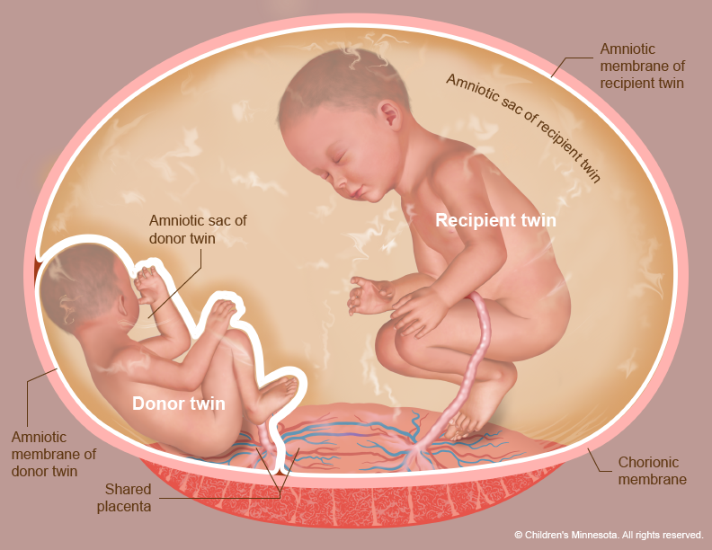
TTTS is a severe complication that affects monochorionic twins, as their shared placenta means that their circulatory systems are connected. Sometimes, the blood vessel connections in the placenta are imbalanced, causing one twin to persistently push blood to or receive extra blood from the other twin. The syndrome is characterized by a low levels of amniotic fluid (oligohydramnios) around one twin (the ‘donor twin’) and too much amniotic fluid (polyhydramnios) around the other twin (the ‘recipient twin’). If untreated, this condition poses considerable risks to both the donor and recipient twin.

Epidemiology

It is estimated that 9 to 15% of monochorionic diamniotic and 6% of monochorionic monoamniotic twin pregnancies will be complicated by TTTS, most commonly presenting between 16 and 26 weeks of gestation. In addition, there is a relatively rare and sudden type of TTTS (intrapartum TTTS) that affects 1.5 to 2.5% of monochorionic pregnancies during labour, regardless of whether the birth was vaginal or a c-section. This type of TTTS results in a rapid and large intertwin blood transfusion from donor to recipient, causing the donor to have too many red blood cells (polycythemia) and the recipient to have too little red blood cells (anemia). The donor twin may also go into shock from the sudden loss of fluid (hypovolemic shock).


Risks of TTTS

TTTS is a severe complication of pregnancy and can result in organ failure and the potential death of either twin. You may also be at higher risk of preterm birth due to the overstretching of the uterus (by the excessive amount of amniotic fluid around the recipient twin) and cervical shortening. Read more about preterm birth in the section above to learn about how that risk is managed!


Diagnosis

If you have a monochorionic twin pregnancy, you will have an ultrasound at least every two weeks throughout pregnancy to allow the detection of TTTS. The first sign of TTTS will be difference in the levels of amniotic fluid between the two babies. More advanced stages include include abnormalities in blood flow to the placenta (detected by ultrasound Doppler studies) and signs of fetal heart failure. In addition, the babies will have a fetal echocardiogram, which is a special ultrasound to detect congenital fetal heart malformations which are more common in monochorionic twins.

Maternal symptoms of TTTS include shortness of breath, premature contractions, and abdominal discomfort, caused from the rapid overstretching of the uterus due to the extra amniotic fluid.

TTTS management will depend on the stage of the syndrome, classified as stages I to V:

  • Stage I: The only finding is differences in the amount of the amniotic fluid between the two twins
  • Stage II: The bladder of the donor twin (the one with the low amniotic fluid) is not visible due to less urine production caused by an even greater amount of blood being transferred to the second twin
  • Stage III: There is evidence of abnormal Doppler blood flow in one or both twins
  • Stage IV: One or both babies are showing signs of severe heart failure (known as hydrops)
  • Stage V: One or both babies passed away in utero


Management

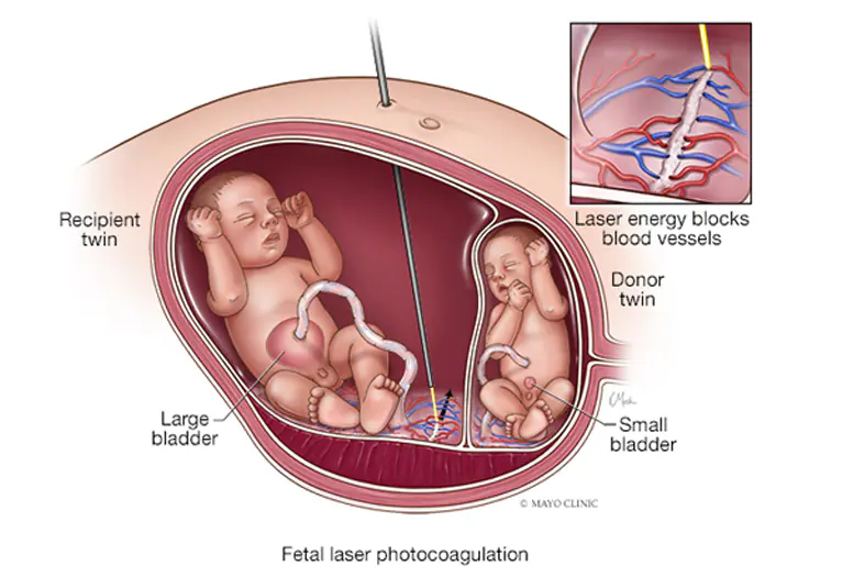
The main procedure used to treat TTTS is fetoscopic laser ablation. It involves inserting a small camera called a fetoscope into the uterus through the mother’s abdomen along with a laser device that is used to destroy (or ablate) the placental blood vessels connecting the two twins. The procedure is done in the operating room under general anaesthesia. Risks of the procedure include preterm prelabor rupture of membranes (PPROM, where the amniotic sac breaks resulting in vaginal leakage of fluid), disruption of the membranes separating the two twins (which can result in entanglement of the umbilical cords of the two babies), and twin anemia polycythemia sequence (TAPS, discussed below).

Source: Mayo Clinic

Twin anemia polycythemia sequence (TAPS)

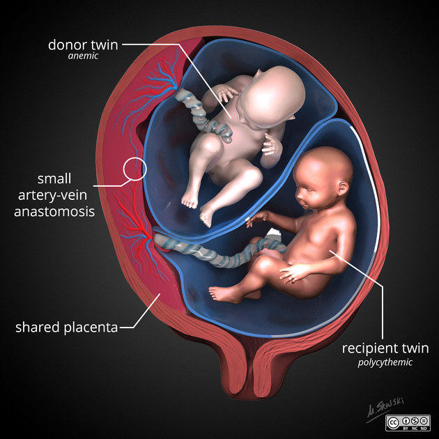
TAPS is a severe complication that affects monochorionic twins, as their shared placenta means that their circulatory systems are connected. You can think of TAPS as a ‘mild form of TTTS’ where the placental connections between the two twins are very small and therefore do not result in differences in fluid (as seen in TTTS) but instead in a very gradual difference in blood hemoglobin (or red blood cells). Red blood cells carry oxygen throughout our bodies to keep our organs functioning via a molecule called hemoglobin. Consequently, this condition is characterized by anemia in one of the twins (less red blood cells and too little hemoglobin to carry oxygen to the organs) and polycythemia (too many red blood cells and too much hemoglobin) in the other.

Source: Radiopaedia


Epidemiology

Spontaneous TAPS is reported in 3 to 6% of monochorionic diamniotic pregnancies. It can also very rarely occur in monozygotic dichorionic pregnancies if there are shared blood vessels between the two placentas. TAPS can also occur post-laser ablation for the treatment TTTS (discussed above) if the laser ablation leaves out small vessels that can allow for the exchange of red blood cells.


Risks of TAPS

The twin with polycythemia has lots of red blood cells which thickens the blood and makes it appear sludge-like. This can cause blood clots and damage the baby’s organs and brain if it stops blood from reaching them. The twin with anemia may develop heart failure. Both twins are at risk of passing away in-utero as a result of this condition. Long-term, there is also a higher risk of adverse neurodevelopmental outcomes.


Diagnosis

Middle cerebral artery-peak systolic velocity (MCV-PSV) is a Doppler measurement that can diagnose anemia and polycythemia in utero. It is based on the idea that, in cases of anemia, the blood flow is more watery (less thick) and will therefore flow faster, the same way water flows faster than oil. Other ultrasound findings include difference in thickness and color of placenta with a clear demarcation between the twins’ territories, a large heart (cardiomegaly) and fluid overload (ascites) in the donor twin, and a certain appearance on the recipient twins’ liver on ultrasound called a starry sky.

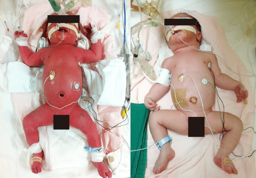
Sometimes the diagnosis is not made until after birth. At that time, TAPS may be suspected if one twin is very pale and the other is red (their respective side of the placenta will show the same colour imbalance). Blood testing can reveal that one twin is anemic and the other is polycythemic, and placental testing may show shared small blood vessels that allow unidirectional flow.

Myung et al. (2016)

Management of TAPS depends on the stage:

  • Stage I: Difference in the MCA-PSV between the two twins is >0.5 MoM (a measurement); with no signs of the babies not doing well
  • Stage II: Difference in the MCA-PSV between the two twins is >0.7 MoM; with no signs of the babies not doing well
  • Stage III: Stage 1 or 2 findings plus heart failure in the donor or recipient twin
  • Stage IV: One or both babies passed away in utero


Management

The treatment options in cases of TAPS involve fetoscopic laser ablation or intrauterine blood transfusion. Your obstetrician will help you make a decision on the best treatment option based on the stage of TAPS, your health, and the health of your babies.

Selective fetal growth restriction (sFGR)

Selective fetal growth restriction (sFGR) occurs when one twin is considerably smaller than the other. The most commonly used definition is that the smaller twin’s estimated weight is below the 10th percentile for gestational age, and that size difference between the twins is greater than 25%.

The size difference is calculated as follows: [WeightOfLargerTwin – WeightOfSmallerTwin] / [WeightOfLargerTwin] x100)


Epidemiology

About 10-15% of monochorionic pregnancies may be affected by sFGR. There is a substantial overlap between sFGR, TTTS, and TAPS, which means that a monochorionic twin pregnancy may be affected by both TTTS and sFGR.


Cause of sFGR

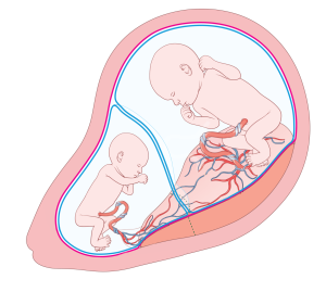
sFGR is thought to happen when the placenta is not shared equally between the two twins (e.g., instead of each twin receiving ~50% of the placental territory, one twin receives only 30%, while the other one 70% of the placental territory). It is thought that the placental connections between the two twins are essential to support and sustain the smaller twin.

Source: TAPS Support

Risks of sFGR

Pregnancies complicated by sFGR are at risk of preterm birth and in-utero demise of the smaller twin (which can also result in in-utero demise of the second twin as well).


Diagnosis and classification

If you have a monochorionic twin pregnancy, you will have an ultrasound at least every two weeks in your second trimester, where the team will estimate the weight of each twin and calculate the size difference between them. As mentioned above, the classic definition of sFGR is that the smaller twin’s estimated weight is below the 10th percentile for gestational age, and the size difference between the twins is greater than 25%. A more recent definition is based on the following criteria:

(i) Estimated fetal weight of less than the 3rd percentile of one twin

OR

At least two of the following:

(i) Estimated fetal weight of less than the 10th percentile of one twin

(ii) Abdominal circumference of less than the 10th percentile of one twin

(iii) Umbelical artery pulsatility index greater than 95th percentile of the smaller twin

(ib) A weight difference between the twins that is greater than or equal to 25%


sFGR is classified into one of three types, based on the by the pattern of the umbilical artery (UA) Doppler in the smaller twin:

Type 1 sFGR: Persistently forward UA end-diastolic velocity without variation in the waveform with normal or elevated resistance. This type is the mildest and has the best outcomes.

Type 2 sFGR: Fixed absent or fixed reversed UA end-diastolic velocity without variation of the waveform in the smaller twin. This type is the most severe form and carries the greatest risk of fetal demise and preterm birth.

Type 3 sFGR: Characterized by UA waveform that has a variable flow pattern that cycles between forward, absent, and reversed flow over a short interval (also called intermittent absent/reversed end-diastolic flow). This is thought to be the result of a larger placental arterial vessel connecting the two twins (large artery-to-artery [AA] anastomosis). While the overall outcomes of cases with type 3 sFGR is considered better than that of type 2 sFGR (since the large AA anastomosis can provide more support to the smaller twin), the outcome in this type is less predictable and there is a greater risk of unpredictable course with sudden demise of one or both twins, as well as greater risk of neurological deficits after birth due to sudden pressure changes through the large AA anastomosis.


Management

The treatment in cases of sFGR depends on the type and severity. It involves close surveillance as an outpatient or on an inpatient basis and early delivery when there are signs of imminent demise of the smaller twin. In more severe cases, other treatment options may be considered including selective termination of the smaller twin or fetoscopic laser ablation. The management of these cases is complex and should be done in a specialized tertiary center experienced in the care of twin pregnancies.

Cord entanglement

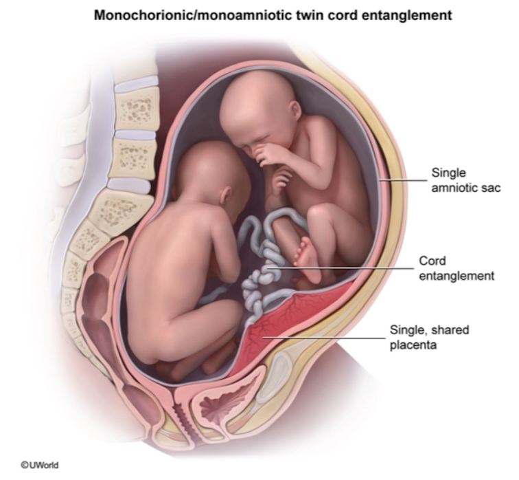
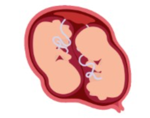
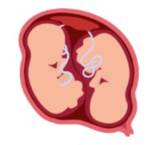
In MC/MA twin pregnancies, there is nothing separating the two babies from each other. Consequently, the twins’ umbilical cords may wrap around each other. This is called . Cord entanglement occurs in almost all MC/MA twin pregnancies. They may occur as early as the first trimester, and incidence increases with increasing gestational age as the babies can move around more and accidentally twist their cords.

In most cases, cord entanglements remain loose and do not cause a problem. However, in some cases, the entanglement might become tight and interrupt blood flow to one or both twins. This can result in a sudden and unpredictable demise of both twins. Studies estimate the overall risk of fetal demise at approximately 18%; however, the risk decreases gradually with increasing gestational age and is lower during the 3rd trimester.

While cord entanglement can be seen on ultrasound, it is not possible to determine whether the entanglement is loose or tight, which makes it very challenging to identify the cases where the risk of fetal demise is increased.

Source: UWorld


Management

 Given that there is no intervention for cord entanglement, the management is based on close surveillance of the babies, with the intention of detecting early signs early signs of tightening that would allow for an urgent delivery by a cesarean section. However, it remains unclear whether the best strategy for close surveillance is through admission to the hospital and observation as an inpatient, versus less frequent monitoring as an outpatient. A recent meta-analysis suggested that the risk of fetal death is lower with inpatient surveillance compared with outpatient management (3% vs. 7.4%).

At Sunnybrook, we offer patients the following management options, highlighting the pros and cons of each option, and make a shared decision regarding the management plan:

  1. Outpatient management – patients will be followed with ultrasound and a non-strress test (NST) x2-3 times per week.
  2. Inpatient management – patients will be admitted to the hospital at approximately 26 weeks to allow for closer fetal surveillance, which includes NST (fetal heart rate monitoring) of 60 minutes x3/day, along with ultrasound x3 per week.

Combination of the two management strategies – some patients may choose to be followed initially as an outpatient and then be admitted to the hospital later in gestation (e.g. at 28 or 30 weeks).

Irrespective of the surveillance strategy chosen, the delivery via c-section is scheduled for 33 weeks, as this gestational age has been shown to offer the optimal balance between the risks of fetal demise (in the case that delivery is deferred) and the small risks of prematurity at that gestational age.

Structural anomalies

MC/MA twins also have a high risk of structural anomalies, up to 25%. It is likely that the splitting of the embryo at such a late stage (after day 8) increases the chance of congenital anomalies. Therefore, all MC/MA twins will undergo an early anatomy scan and fetal echocardiogram (before 14-15 weeks’ gestation) to allow for a timely detection of such anomalies.

Conjoined twins

Conjoined twins are twins whose body parts have fused together. Conjoined twins occur when the single embryo splits at an even later stage than in the case of MC/MA twins, at a point where fetal structures have already started to develop. As a result, the two twins will have to share those body parts that had already started developing by that time. There is great variation in the degree and the type of fusion.


Epidemiology

Conjoined twins are rare, with an incidence of approximately 8 out of 100,000 births. However, it is likely that their incidence is underestimated, given that many of these pregnancies result in early miscarriage before the diagnosis of conjoined twins can be made.


Risks of conjoined twins

There is a high risk of stillbirth with conjoined twins, as well as congenital abnormalities. In addition, conjoined twins born alive often require. For example, separation surgeries for thoraco-omphalopagus conjoined twins (joined at the thorax and anterior abdominal wall, most common with approximately 75% of conjoined twins) may involve separation of the sac around their hearts and their gastrointestinal tracts. Tissue expanders can be used to stretch the existing skin to later have enough for both twins. The extent of surgery and type are unique to the specific twin pair and which of their body parts and organs are fused.


Diagnosis

Conjoined twins can be seen on ultrasound, which you will be undergoing routinely during your pregnancy. Additional imaging such as color Doppler, fetal echocardiography, 3D ultrasound, and fetal MRI may be used to confirm the diagnosis, clarify anatomy, and help with decision-making (e.g. for surgical planning).


Management

Many specialists are involved in the care of these pregnancies. This includes your obstetrician, who is also an expert in maternal-fetal medicine, a pediatric surgeon, a neonatologist, and a radiologist.

Lifestyle

Can I exercise?

There isn’t a lot of evidence to link restricting exercise and physical activity, or leaving work early, with preventing preterm labour in women pregnant with twins. However, some physicians recommend that women pregnant with twins should restrict physical exercise after 28 weeks.

We routinely monitor the length of your cervix during pregnancy. Having a cervical shortening increases the risk of preterm birth. In cases in which the cervix is found to be short or in the case of preterm contractions we will advise that you restrict physical activity.

Are there good online calculators for my pregnancy and determining potential due dates?

Check out this calculator from perinatology.com

Can I have sex?

Can I have sex while pregnant with twins?

Intimacy is an important part of your relationship with your partner. Sexual activity is not discouraged in twin pregnancies, unless there are complications that your healthcare provider has discussed with you.

Sunnybrook’s Twins Clinic recommends against sexual intercourse if you have both a short cervix and are pregnant with twins, especially in cases with severe or early-onset cervical shortening.


To understand a bit more about this issue, we have compiled available evidence on sex in twin pregnancies below. This information is taken from a book chapter our team has written on the topic.

Twin and triplet pregnancies have been associated with an increased risk for complications, primarily preterm birth. The increased risk for preterm birth may have individuals pregnant with twins and their providers concerned that intercourse may prompt preterm labor or delivery. The anxiety may be even greater in women with twins who are thought to be an increased risk of preterm birth, such as those with a short or dilated cervix.


Common Questions:

  1. What are the possible effects of prostaglandins (hormones that play a role in induction of labour and uterine activity) in semen on preterm labour?
  2. What are the possible effects of mechanical stimulation (pressure during intercourse) of the cervix during intercourse on preterm labour?
  3. What are the possible effects of uterine contractions during female orgasm and preterm labour?

The Evidence:

  1. Prostaglandins
    • Prostaglandins (PGs) have been shown to be present in the amniotic fluid, placenta, myometrium, and blood at the start of labour, and have also been shown to induce labour and stimulate uterine activity.1,2
    • There have been several studies in singletons on the topic of coitus and the onset of labour, all of which found no relationship between coitus at term and onset of labour.3–6 These studies had major limitations, thus there is no strong evidence about the effect of semen on labour onset.
    • No studies have been done on this topic in twin pregnancies.
  1. Mechanical stimulation
    • Another concern related to coitus in pregnancy is mechanical stimulation of the cervix or lower-uterine segment, which may release prostaglandins.
    • There are no studies that examine endogenous prostaglandin levels after intercourse, nor are there studies comparing condom use to coitus without a condom (which may aid in separating mechanical stimulation causing release of prostaglandins, or prostaglandins from semen). In addition, the cervix and lower-uterine segment in low-risk pregnancies seems to be able to tolerate the impact of penetration during coitus.
    • No studies have been done on this topic in twin pregnancies.
  2. Female orgasm
    • Researchers have observed similarities between the uterine contractions and release of oxytocin during female orgasm and uterine contraction patterns of labour, raising concerns that female orgasm may elicit contractions and stimulate preterm labour.
    • All the research on this topic has been performed in singletons, and most of the studies were published in the 1970s. Despite this, one study performed in 2001 concluded that sexual activity was associated with a reduced risk for preterm birth.7 It is important to note that even strong uterine contractions due to orgasm are likely not as strong or long-lasting enough to induce labour.
    • No studies have been done on this topic in twin pregnancies.

Is there evidence from Twin Pregnancies?

  • There have only been two published, peer-reviewed research studies on the topic of sexual activity and twin pregnancies.8,9 Both studies did not find a relationship between sexual behaviour and complications such as preterm birth in twin pregnancies. In addition, a recent survey of 90 Canadian Maternal-Fetal Medicine specialists found that 97% (87/90) of specialists agree that sexual activity does not need to be avoided in an uncomplicated twin pregnancy.10

If you experience discomfort related to sex during your twin pregnancy, avoid lying on your back during intercourse after the fourth month of pregnancy. Try different positions to reduce pressure and use a personal lubricant to help with dryness.

If you notice pain, bleeding, abnormal discharge, or contractions after sex, we recommend you call your healthcare provider.

And remember, intimacy goes beyond sexual intercourse—it is not just sex. Intimacy is about closeness, about being together, and about creating and maintaining a relationship.


If you have any questions about sexual activity during your twin pregnancy, do not hesitate to ask your healthcare provider at your next prenatal visit.

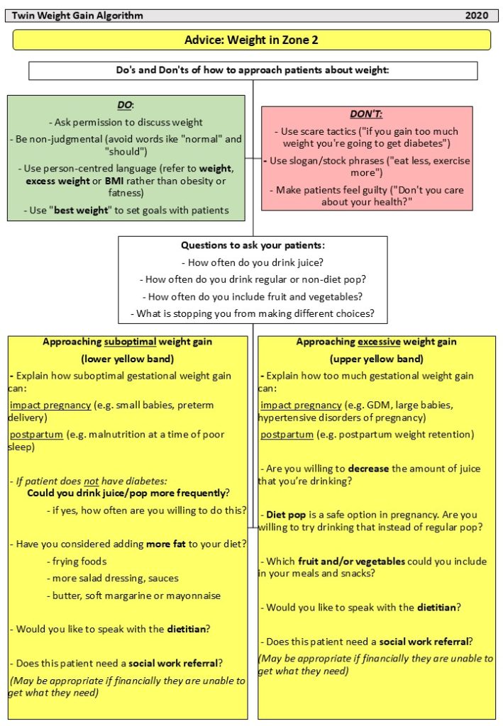
How much weight should I gain?

Weight Gain in a Twin Pregnancy

One of the first questions many pregnant individuals have is: “How much weight should I gain while pregnant?”


Your weight gain during pregnancy is made up of weight gain related to YOU (increase in body water, increase in size of the breast tissue and your uterus, and increase in fat mass) and YOUR BABIES (placenta, amniotic fluid, and, of course, your twins).


Too much or not enough weight gain may increase the risk of some conditions in pregnancy. Gaining too much weight while pregnant might lead to gestational diabetes and preeclampsia. On the other hand, gaining too little weight might lead to preterm birth and might slow down the growth of the babies.


When you’re pregnant with twins, you are already at a higher risk for the complications described above (preterm birth, gestational diabetes, preeclampsia, and fetal growth restriction). Therefore, meeting weight gain targets during pregnancy and reaching the increased nutritional demands of a twin pregnancy may help decrease the risk of these complications.


We’re here to help!


Existing guidelines provide detailed recommendations on the ideal weight in SINGLETON pregnancies. However, when it comes to TWIN pregnancies, professional societies provide very little guidance on the ideal amount of weight gain. Therefore, we conducted a series of studies in the Twins Clinic at Sunnybrook on the weight gain in twin pregnancies.


  • Lipworth H, Amir K, Arruda I, Fockler M, Jung E, Po L, Barrett J, Melamed N. A new care pathway to optimize gestational weight gain in twin pregnancies. Am J Obstet Gynecol MFM. 2023 Aug;5(8):101018. Click here for full paper.
  • Lipworth H, Barrett J, Murphy KE, Redelmeier D, Melamed N. Gestational weight gain in twin gestations and pregnancy outcomes: a systematic review and meta-analysis. BJOG. 2022 May;129(6):868-879. Click here for full paper.
  • Lipworth H, Melamed N, Berger H, Geary M, McDonald SD, Murray-Davis B, Murphy KE,  Redelmeier DA, Yoon EW, Barrett JFR, Ram M; Diabetes, Obesity, and Hypertension In Pregnancy Research Network Investigators. Maternal weight gain and pregnancy outcomes in twin gestations. Am J Obstet Gynecol. 2021 Nov;225(5):532.e1-532.e12. Click here for full paper.


How do I track my weight gain?

To meet this knowledge gap, the team at the Sunnybrook Twins Clinic has conducted developed a series of studies to determine the optimal weight gain your weight gain during pregnancy (see above).


The recommended weight gain in twin pregnancies is based on your pre-pregnancy body mass index (BMI), which is calculated as your weight (in kg) divided by squared height (in m):  You can calculate your pre-pregnancy BMI here.


Once you know what BMI category you are in, use the following table to guide your weight gain throughout pregnancy:


BMI Group

Pre-pregnancy

Recommended total weight gain at 37 weeks Recommended weekly weight gain
Underweight or normal weight
(BMI<25.0 kg/m2)
16.8-24.5 kg

or
(37-54 lbs)

0.45-0.66 kg per week

or
(1.00-1.46 lbs per week)

Overweight
(BMI 25.0-29.9 kg/m2)
14.1-22.7 kg

or
(31-50 lbs)

0.38-0.61 kg per week

or
(0.84-1.35 lbs per week)

Obese
(BMI≥30.0 kg/m2)
11.3-19.1 kg

or
(25-42 lbs)

0.31-0.52 kg per week

or
(0.68-1.14 lbs per week)

BMI, body mass index


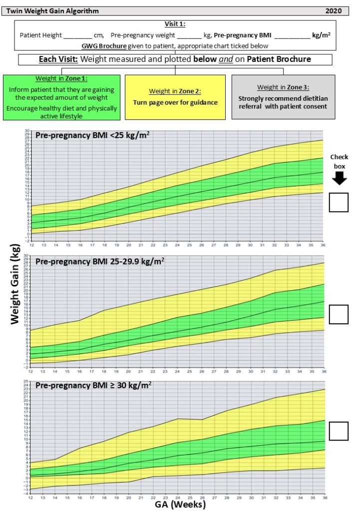
We also developed a tool that can help you track your weight gain during pregnancy. The weight gain charts can be seen below, and you can use this link to use the calculator we developed for that purpose.


In the Twins Clinic, we will monitor your weight gain together throughout pregnancy. In cases of insufficient or excessive weight gain, we will offer you a referral to our dietitian to discuss ways to optimize weight gain.


Nutrition

When pregnant with twins, trying to get a balanced diet with enough protein, carbohydrates, calcium, and energy is important for your and your babies’ health.


Protein

Protein helps with babies’ growth and supports your needs while carrying them. We developed a calculator to help you calculate the protein requirements, specifically in twin pregnancies.

  • Include protein with every meal. Protein is found in:
    • Eggs
    • Fish – choose low mercury options like salmon, rainbow trout or sardines
    • Poultry (chicken, turkey)
    • Meat
    • Legumes (beans, lentils, chickpeas)
    • Greek yogurt
    • Cheese, cottage cheese
    • Tofu
    • Textured vegetable protein (e.g., veggie burgers, veggie dogs)
    • Nuts, nut butters (e.g. peanut butter)

A woman pregnant with twins needs an extra 50 grams of protein each day compared to someone pregnant with one baby.

  • Those extra 50 grams would be equal to:
    • 220 grams (8 ounces) of meat,
    • 8 large eggs, or
    • about two and a half cups of cooked lentils
  • Keep in mind that this is on top of the usual amount needed for pregnancy.
    • A 60 kg (132 lb) person with a twin pregnancy would need about 116 grams of protein per day.


Carbohydrates

Carbohydrate needs are also higher in twin pregnancy. We recommend aiming for a minimum of 208 grams of carbohydrate daily to help with babies’ development, increase your weight gain, and to prevent ketones (a chemical your body makes when you don’t get enough carbohydrates).

  • This is equal to:
    • 6 ½ slices of whole wheat bread,
    • 4 cups of rice,
    • 5 cups of cooked pasta,
    • or 7 ½ chapatis.


Calcium

Getting enough calcium is important to help with growth and maintenance of bones and teeth for you and babies. It’s also important for helping to control blood pressure during pregnancy.

  • All pregnant women should include 3-4 servings of calcium daily to meet their needs.
  • Prenatal vitamins usually have 1 serving and the rest can come from food.
  • Mix and match the calcium-rich foods that you enjoy to get enough.
  • Each of the following is considered 1 serving of calcium:
    • 1 cup of milk or fortified milk alternative (e.g. soy beverage, rice beverage, almond, oat beverage)
    • 1/3 cup of cubed pasteurized cheese
    • 1 cup of yogurt


Nausea and Food Intake

It can be challenging to get enough food in if you are feeling nauseated or if your babies are not leaving you a lot of room in your stomach to eat. A couple of tips to help you get enough to eat include:

  • Have small, frequent meals and snacks with both carbohydrates and protein, for example:
    • Fruit and cheese
    • Greek yogurt and cereal
    • Hummus and pita
    • Sandwich with a cup of milk
    • Crackers with peanut butter

If you are finding it difficult to gain weight or get enough to eat, ask your care provider for a referral to a registered dietitian.

Protein and pregnancy

Getting enough protein during pregnancy will help support growth and development of your baby/babies while maintaining your muscle mass. You can use this link to access a tool we developed that can help you track your weight gain during pregnancy. Use the examples below to help meet your protein needs.


Poultry and Meat

A 3 ounce (85 gram) portion of meat or poultry is about the size and thickness of the palm or your hand and has the amount of protein listed below each image.


Fish and seafood

Some fish or seafood can be measured by comparing it to the palm of your hand. A 3 ounce (85 gram) cooked portion of fish or seafood is about the size and thickness of the palm of your hand. Other fish or seafood may be easier to portion using measuring cups or counting the pieces. The portions are listed here as is the amount of protein.


High protein dairy products
Dairy products may be easier to portion using measuring cups or spoons. The portions are listed here as is the amount of protein.
Other high protein foods

Other high protein foods may be easier to portion using measuring cups or spoons. The portions are listed here as is the amount of protein.


Labour and Delivery

How should I give birth - vaginal delivery versus caesarean section?

The answer about how you will deliver your twins depends on several factors. The primary factor is the way the first twin to come out is positioned within your uterus. When the first twin is in a breech presentation, delivery should be via C-section.


When the first twin has their head down, the best way to deliver has been a matter of debate between obstetricians over the years. Some believe C-section is safer. Others recommend vaginal delivery only if the second twin is also positioned with the head down, and C-section if the second twin is in the breech position. Many other obstetricians believe that vaginal delivery is the best choice when the first twin is with the head down, regardless of the position of the second twin.


A large randomized controlled trial, led by Dr. Barrett (the Twin Birth Study) was conducted specifically to address this point. The trial provided evidence that as long as the first twin is with the head down, vaginal delivery is as safe as a C-section, and this is the approach that is taken at Sunnybrook.


A vaginal twin birth is somewhat different that the delivery of a single baby. The delivery of the first twin is essentially similar to that of a normal delivery in a singleton pregnancy. The main difference is that once the first twin is out, there is a small risk of complications for the second twin, including cord prolapsed, detachment of the placenta (placental abruption) due to the sudden decompression of the uterus after the delivery of the first twin, or change in the position of the second twin. Because of that, there is a risk of approximately five per cent that an urgent C-section will be needed for the second twin. For that reason, vaginal twin birth always takes place in the operating room, with the team being set up for urgent C-section if needed.